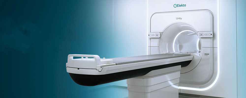
The magnetic resonance-guided linear accelerator (MR LINAC) combines MRI with radiation treatment to target and cure malignancies. Doctors may now alter radiation therapy in real time with improved soft tissue resolution and deliver it more correctly and effectively than ever before, thanks to MRI guidance. The MR Linac system combines two technologies—an MRI scanner and a linear accelerator—to precisely pinpoint tumours, shape X-ray beams in real time, and administer radiation dosages correctly, even to moving tumours.
If you are a candidate for MR LINAC therapy, your medical team will create a treatment plan for you. They will also implement quality assurance methods to verify that each therapy is delivered in the same manner. An MR-LINAC simulation will be performed on you prior to the treatment.
Doctors employ MR LINAC to treat cancer patients. It targets the patient's tumour using high-energy x-rays. These therapies may be designed by doctors and physicists such that the radiation eliminates the cancer cells while preserving the surrounding normal tissue.

The MR LINAC employs continuous MRI to monitor the movement of soft tissue and organs. This allows doctors to detect tumour migration and subsequently correction during therapy. This is beneficial for tumours in the lung, prostate, intestine, and bladder that move around a lot.
Soft tissues are better visualised with MRI than with x-rays or other imaging. Treatment is safer, with fewer possible adverse effects, and a bigger dose can be used to attain the aim. This reduces the possibility of harming vital organs around the tumour.
The MR LINAC is equipped with innovative software that allows your doctors to modify your radiation treatment plan based on what they see on a daily basis.
MR LINAC represents a significant technical advancement in cancer therapy. To offer therapy, it combines the imaging capacity of an MRI with that of a linear accelerator. To minimise interference, the treatment team divides the MR LINAC's magnetic field to make room for the linear accelerator. Radiation can now travel through the gap. MRI can then produce pictures that are free of distortion.
MRI, or magnetic resonance imaging, produces comprehensive pictures of organs and tissues throughout the body without the use of x-rays. Instead, MRI creates pictures using a high magnetic field, radio waves, quickly changing magnetic fields, and a computer. These photos can reveal the presence of an injury, disease process, or aberrant state.
The patient rests within the MR system or scanner for the MRI test, which is generally a huge donut-shaped device that is open on both sides. The strong magnetic field aligns atomic particles known as protons, which occur in water-containing human tissues. The applied radio waves subsequently interact with these protons to generate signals that are picked up by a receiver within the MR scanner. The rapidly shifting magnetic fields are used to characterise the signals. Cross-sectional pictures of tissues are created as "slices" by computer processing, which the radiologist may see in any direction.
An MRI exam is not painful. There is no documented tissue damage caused by electromagnetic radiation. During the operation, the MR system may emit loud tapping, knocking, or other noises. To avoid issues caused by the noise created by the scanner, the radiation therapist will supply earplugs. Yashoda Hospital staff will always keep an eye on you. You will be able to speak with the radiation therapist via intercom or other means.
The linear accelerator accelerates electrons using microwave technology (similar to that used in radar) in a section of the accelerator known as the "wave guide." When the electrons hit a heavy metal target, high-energy x-rays are produced. As they depart the MR LINAC, these high-intensity x-rays are shaped to correspond to the shape of the patient's tumour.
To shape the beam, a multileaf collimator built within the machine's head is commonly employed. The patient is positioned on a movable therapy couch. Lasers are used by the radiation therapist to ensure that the patient is in the appropriate posture. The treatment couch may be moved in a variety of directions, including up and down, right and left, and in and out. The beam emerges from a section of the accelerator known as a gantry, which may spin around the patient. By rotating the gantry and adjusting the treatment couch, the system can administer radiation to the tumour from a variety of angles.

Internal inspection methods are also included in modern linear accelerators. These mechanisms prevent the machine from turning on unless all of the stipulated treatment conditions are satisfied.
The radiation therapist at Yashoda Hospital monitors the patient on a closed-circuit television monitor during treatment. In the treatment room, there is also a microphone so that the patient may communicate with professionals if necessary.
The safety of the personnel operating the linear accelerator is also critical. The linear accelerator is housed in a chamber with walls made of lead and concrete. This protects against high-energy x-rays. This implies that the x-rays are not visible to anyone outside the room. From outside the treatment chamber, the radiation therapist must activate the accelerator.
The risk of unintentional exposure is quite minimal because the accelerator only produces radiation when it is turned on.
Yashoda Hospital is a medical innovation leader, most notably for its revolutionary MR Linac therapy. Dr. Ravi Suman, a prominent neurosurgeon known for his expertise and trailblazing contributions to the discipline, is at the forefront of this innovative technique. Dr. Ravi Suman has revolutionised the treatment of brain tumours as one of the country's first few neurosurgeons to master this complex surgery. His exceptional talent and dedication to advance medical knowledge have allowed patients to benefit from cutting-edge technologies, providing fresh hope and improved outcomes in the fight against brain tumours. Yashoda Hospital's drive to push the frontiers of healthcare with novel therapies like the MR Linac demonstrates its passion for providing patients with superior and progressive care.
Generally, MR Linac is a safe procedure. In some cases, a contrast material named gadolinium is given to the patient, which can rarely cause allergic reactions. Before receiving the contrast agent, talk to the radiologist if you have kidney failure, kidney transplant, kidney disease, liver disease and related conditions.
MR-Linac offers two diverse technologies into one. The MRI targets tumor while linear accelerator delivers high dose radiation to kill cancer cells. This technology allows oncologists to check for tumor in real-time and avoid harming healthy tissue.
The following cancers can be treated through MR Linac:
People undergoing MR-Linac may feel the following side effects: